mucoderm® is characterized by a porous collagen structure, visualized by the innovative synchrotron-based X-ray tomography: Synchrotron-based X-ray tomographic microscopy for visualization of three-dimensional collagen matrices.
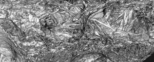
Synchrotron-based x-ray image showing the porous structure of mucoderm®
This unique structure makes mucoderm® an ideal scaffold for ingrowth of blood vessels and cells and promotes fast tissue integration and revascularization. Tissue integration and degradation were analyzed in an animal (rat model) study of Prof. Daniel Rothamel4. After only two weeks, mucoderm® shows an extensive ingrowth of blood vessels as well as an inflammation-free healing with superficial cell invasion. In the following four to eight weeks, a continuous degradation with increasing homogeneous cell distribution can be observed. After eight weeks, still 20% of the original matrix volume is available as a scaffold for the formation and reorganization of connective tissue. After twelve weeks, mucoderm® is almost completely replaced by newly formed connective tissue.