Bone augmentation with the shell technique
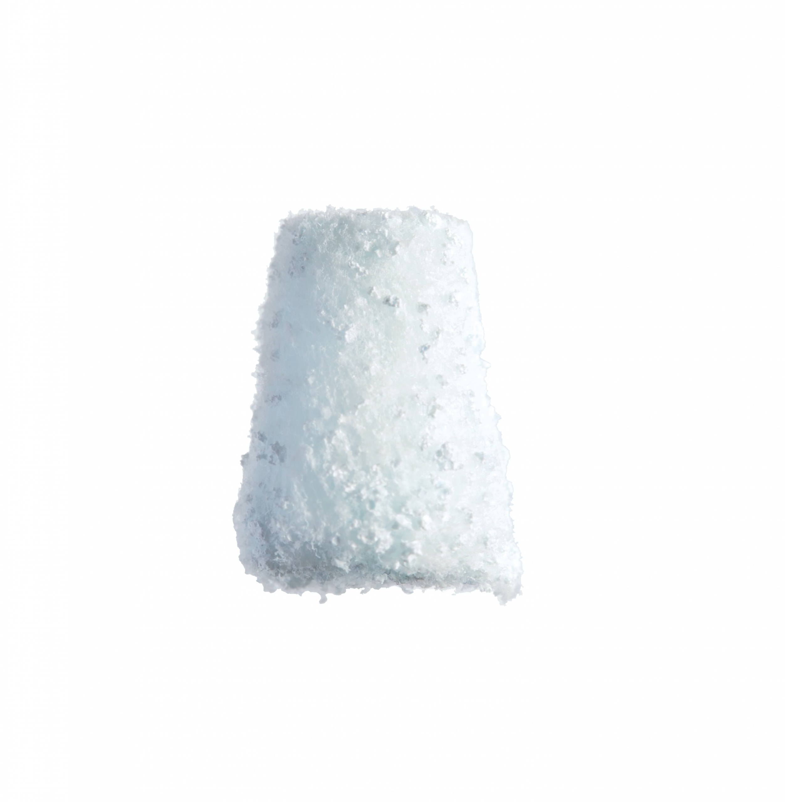
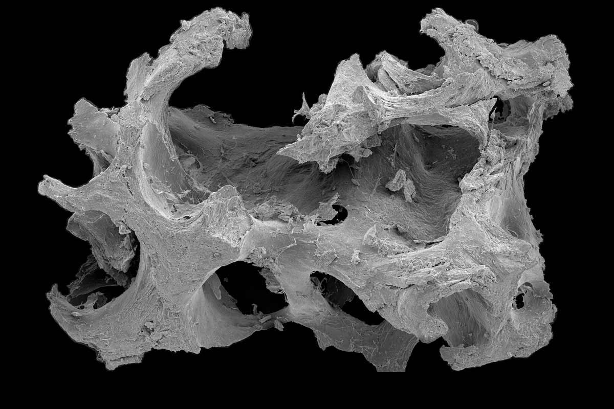
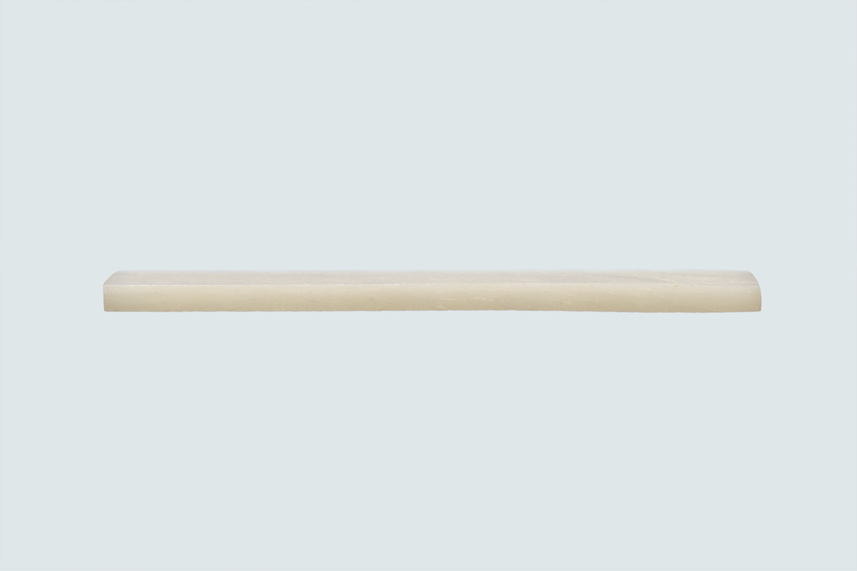
maxgraft® cortico is an allogenic bone graft from human donor bone, which is prepared by Cells+Tissuebank Austria in a special purification process (Allotec® process). maxgraft® cortico is a thin, stable cortical plate specially developed for the shell technique. The shell technique creates a biological container which can be filled with particulate bone substitute material, facilitating revascularization and migration of bone-forming cells into the defect zone for rapid bone regeneration1.
SHORTER TREATMENT TIMES
ALTERNATIVE TO AUTOLOGOUS PLATES
STABLE CORTICAL BONE SCAFFOLD
Allogenic Shell Technique
High Patient Acceptance – Shorter Treatment Times
Properties & Advantages
Indications
Privacy Policy
Valid Alternative to Autologous Plates
maxgraft® cortico spares the patient the possible source of complications of autologous bone harvesting, the donor site morbidity, as well as the associated pain, which is often greater than with the augmentation itself. The practitioner is also spared the need to divide the bone block and the time-consuming thinning of the bone platelets.
Osteoconductive Space for Bone Regeneration
The shell technique creates a biological container between the local bone and the cortical plate. This free space can be filled with the surgeon’s preferred particulate bone substitute material, such as maxgraft® granules or autologous bone chips. The osteoconductive characteristics of maxgraft® allows natural remodeling.
Stable Cortical Bone Scaffold
Postoperatively, maxgraft® cortico is primarily integrated. Since the bone plate consists of cortical bone, it is not rapidly resorbed by the body but gradually remodeled. New vital bone forms directly on the side of the allogenic plate facing the local bone. Compared to autologous bone plates, maxgraft® cortico shows comparable, sustained stability.
Rehydration
Rehydration of maxgraft® cortico is recommended. Rehydration of maxgraft® cortico (10 minutes in saline solution) has been shown to increase flexibility and improve the fracture strength of the plate.
Origin
maxgraft® is processed bone graft substitute material from human donors. maxgraft® cortico is completely mineralized cortical bone and originates from the femoral diaphysis of post mortem donors. Procurement is standardized according to a predefined procurement protocol and is performed by certified procurement centers. All tissue donations are made only after the donor has given written consent. In addition, the health status of each potential donor is assessed in advance as part of a risk analysis and the donor is then selected on the basis of strict exclusion criteria.
Safe and Sterile
After a thorough analysis of the donors’ medical history, the high safety of maxgraft® is ensured by a series of rigorous serological tests in combination with the Allotec® purification process of the C+TBA and final radiological sterilization. C+TBA is certified as a tissue procurement facility and tissue bank according to §19 and §22 of the Austrian Tissue Safety Act.
NOVAMag® membrane
Resorbable Magnesium Membrane
Jason® membrane
Native Pericardium GRB/GTR Membrane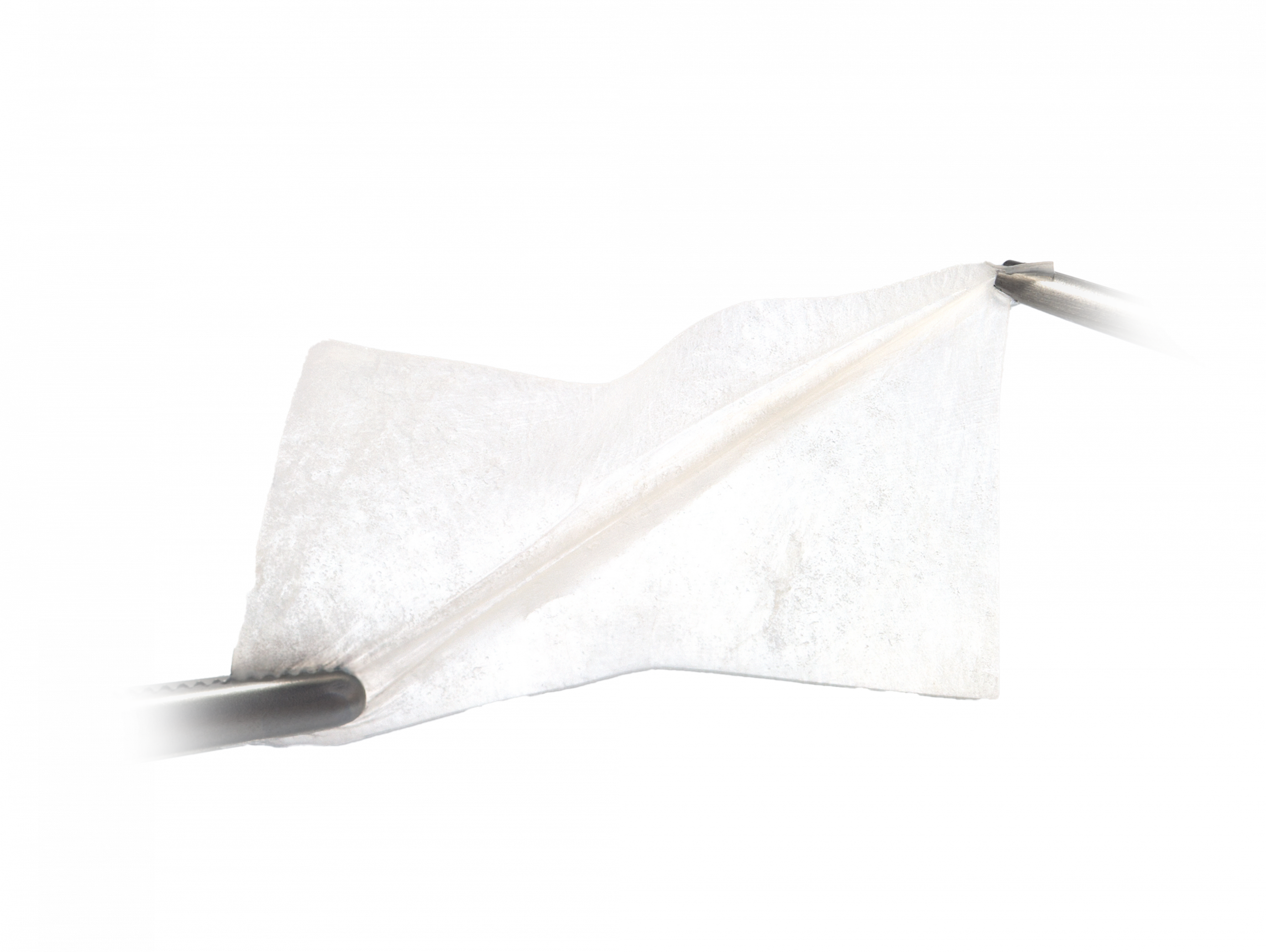
maxgraft® granules
Processed Human Allograft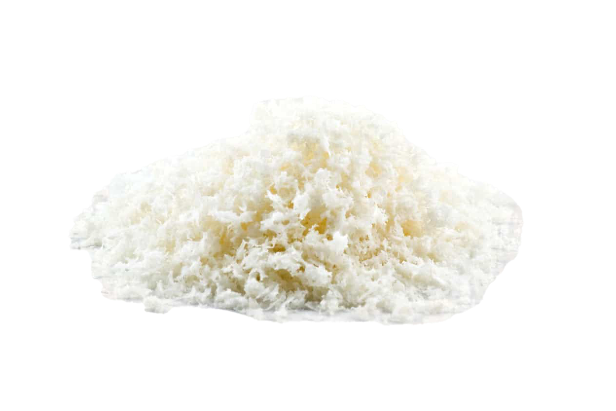
maxgraft® blocks
Processed Human Allograft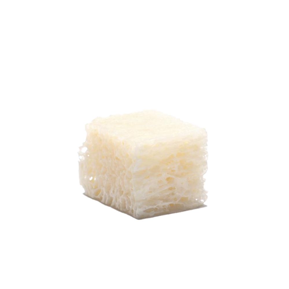
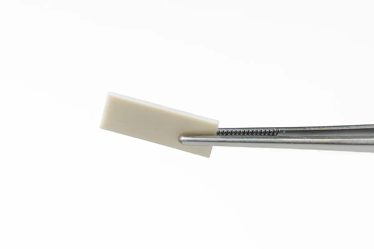
maxgraft® cortico
Bone augmentation with the shell technique
maxgraft® bonebuilder
Customized Allogenic bone block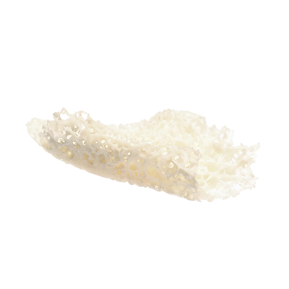
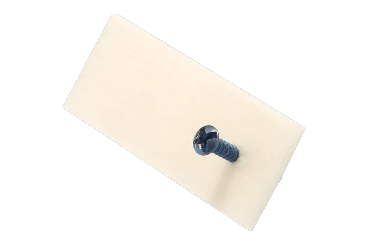
NOVAMag® fixation screw
Resorbable magnesium screws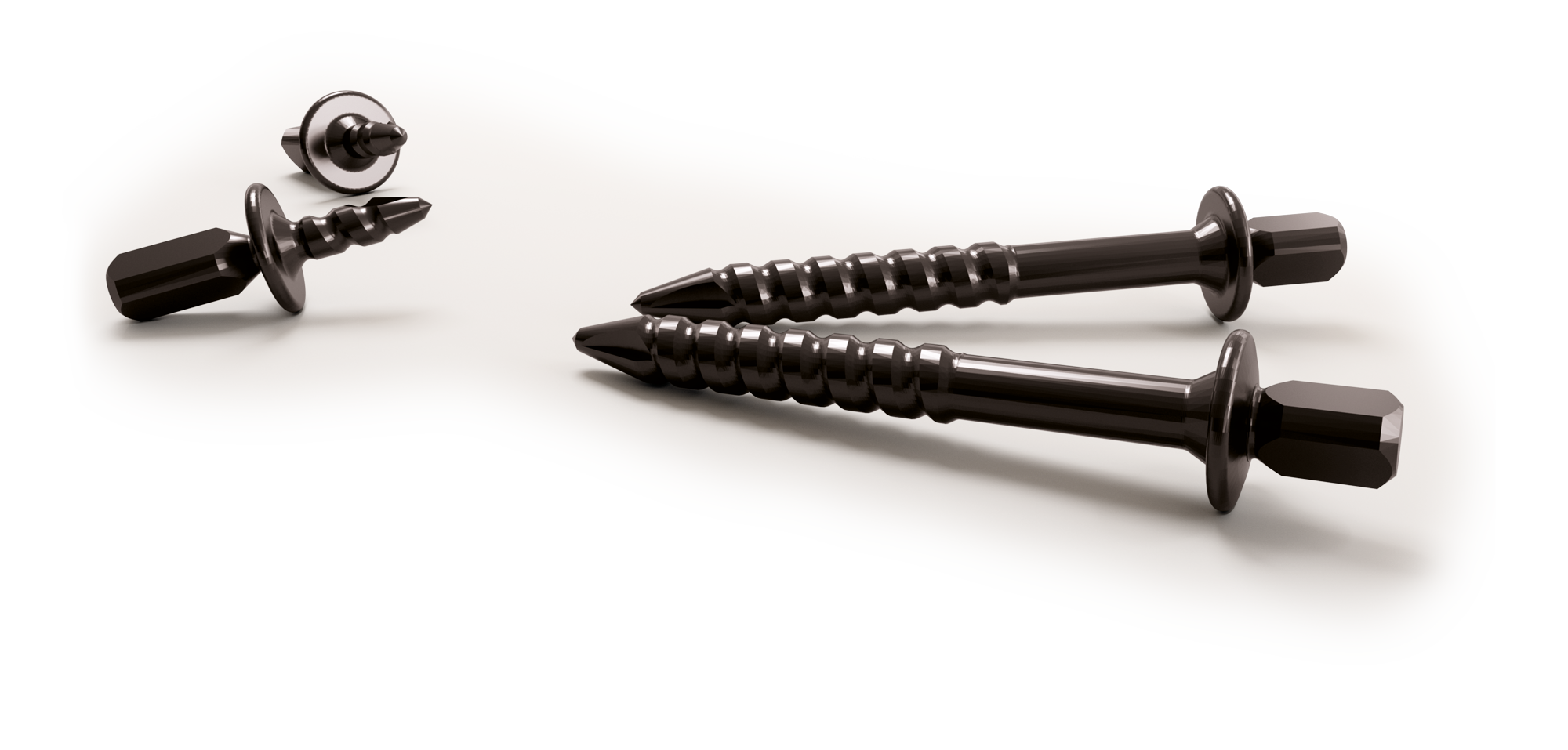
mucoderm®
Acellular dermal collagen matrix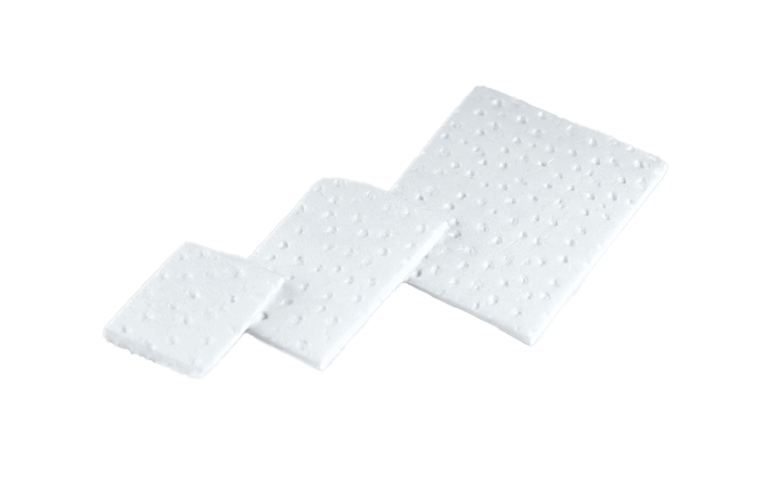
collafleece®
Resorbable collagen for wound management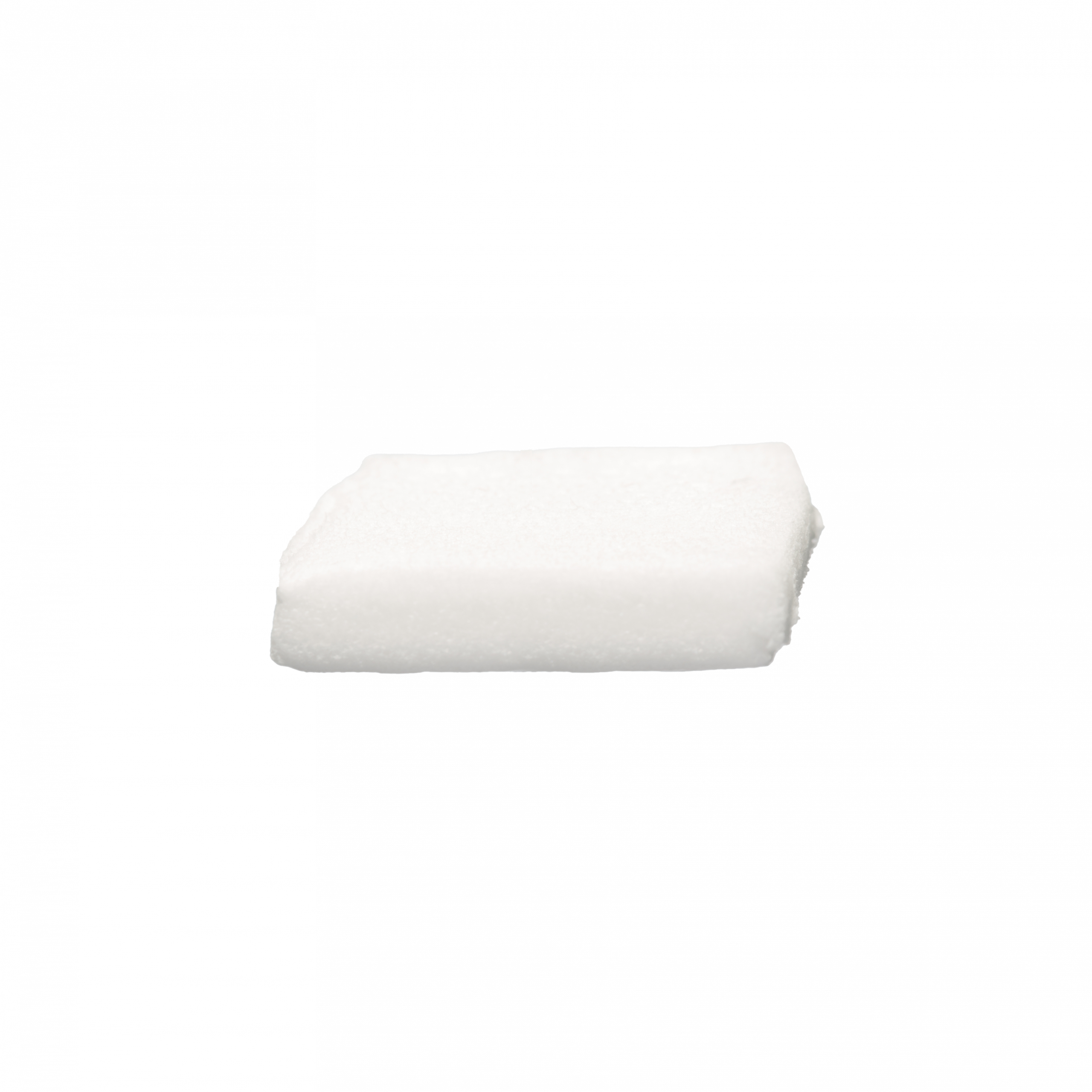
collacone®
High-Density PTFE Barrier Membrane